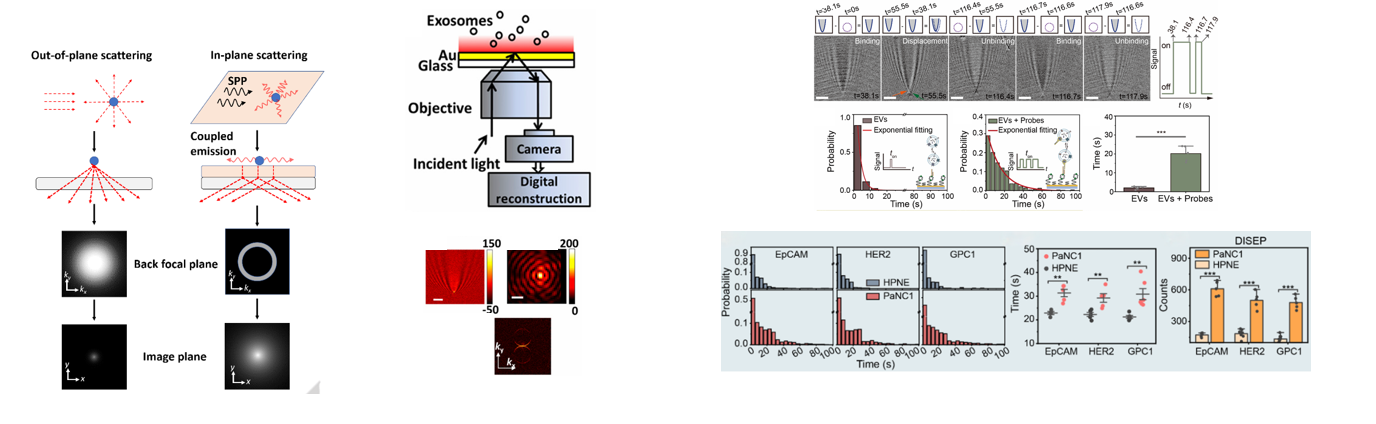
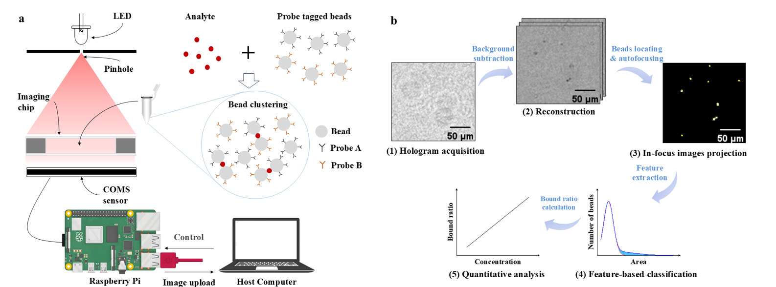
Yu's Lab
  • Home
  • Research
  • Publications
  • News
  • Gallery
  • Contact Us
  • Team
    PI
    Members
    Alumni
    External Collaborator
  • Postdoc Positions Available/本课题组常年招收博士后,待遇优厚, 有意请联系!
    • In-vitro diagnosis

    • ① Exosome detection

        Supermeres are extracellular nanoparticles with a diameter of less than 30nm that are abundant in various body fluids. They contain a rich variety of highly heterogeneous cancer biomarkers and can be used in liquid biopsy for disease diagnosis and early screening. Moreover, they can be extracted from body fluids non-invasively or minimally invasively, offering significant advantages over tissue biopsy.
        The InS imaging technology developed by our group can capture extremely small supermeres particles on an improved silicon nitride chip to achieve imaging and count the binding quantity. By taking advantage of the difference in binding time between specific and non-specific binding, the concentration of specific cancer biomarkers in the sample can be analyzed to achieve cancer diagnosis.

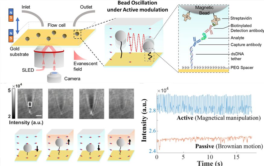
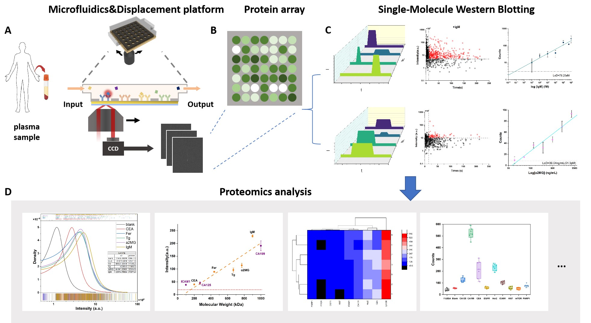
    • ② Ultra-sensitive immunoassay


        Single-molecule detection technology has ultra-sensitive detection capabilities, but non-specific binding can lead to false positives, especially in complex biological samples, where the analysis accuracy is relatively low. Therefore, we have proposed a method of magnetic regulation dynamic sensing to identify the specific binding of individual analytes through periodic modulation.
        In addition, we have proposed digital Western blotting technology. Through dual detection of quality screening and immune verification, it can achieve femtomol-level trace marker detection, which is expected to assist in the early diagnosis of diseases such as cancer

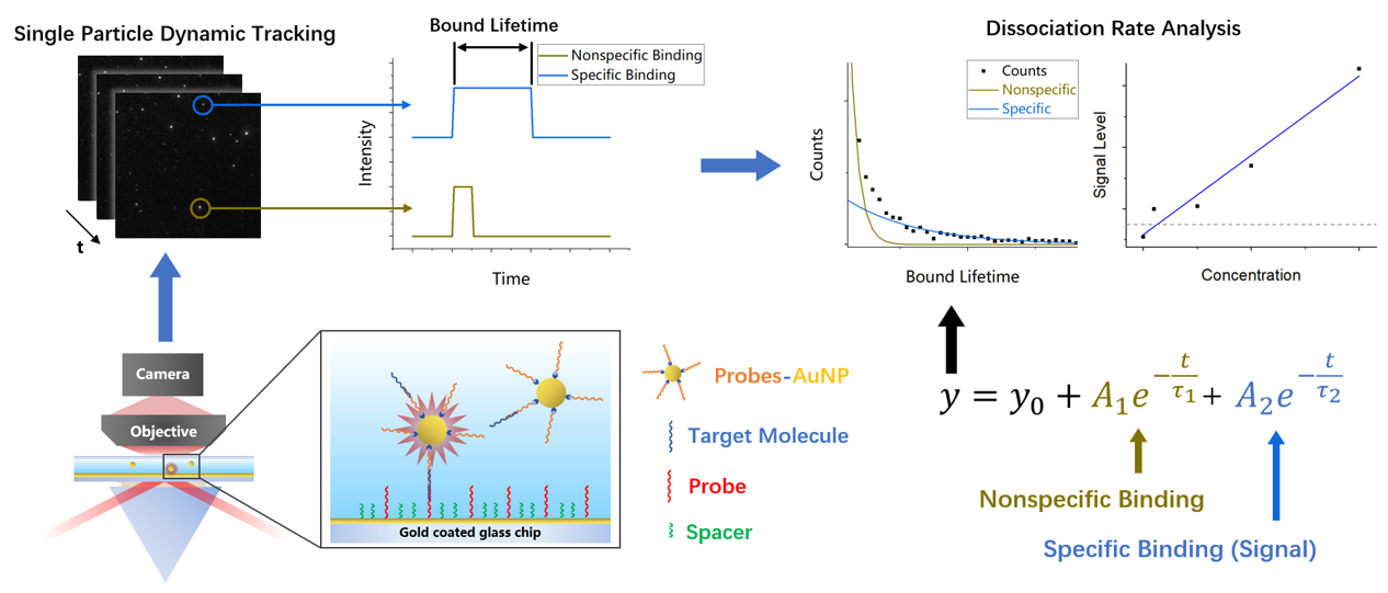
    • ③ Molecular binding kinetics

        Protein interactions drive essential biological processes, and precise measurement of their kinetic parameters is crucial for drug screening, target validation, and biomarker discovery. Traditional labeling methods like fluorescence and radioisotopes disrupt native protein conformations and only provide endpoint data. While label-free techniques like SPR and BLI can detect binding kinetics, their sensitivity is fundamentally limited by the low mass of small molecules. To address this, our research focuses on developing novel SPR-based sensing technologies with enhanced sensitivity for quantifying protein-small molecule interactions, thereby advancing both fundamental research and drug development.

    • Organoid analysis

    •   Organoids are multi-cell clusters formed by three-dimensional culture of stem cells or tumor cells in vitro. Its growth process mimics the process of human development or in vitro organ regeneration. Tracking and analyzing the formation process of organoids can help study the potential mechanisms of human development and organ regeneration, and promote research in basic biology.
        Based on multi-angle illumination lens-free imaging technology, we have designed a three-dimensional imaging system with a large measurement range, single-cell resolution, label-free, small volume (placed in an incubator), and fast imaging speed. Currently, we are dedicated to multimodal organoid analysis, including morphological and membrane protein imaging

    • Label-free Imaging

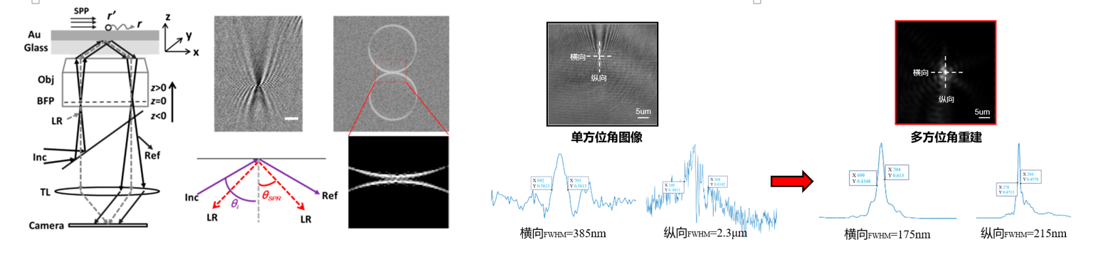
    •   Traditional plasma resonance microscopy (SPRM) is widely used in the fields of biology, chemistry, etc. due to its advantages such as no labeling and high sensitivity. However, the classic "parabolic" images obtained by traditional SPRM have problems of insufficient spatial resolution and anisotropy.
        To address the drawback of insufficient spatial resolution of SPR "parabolic" images, our group, based on its anisotropic characteristics, have optimized the imaging optical path by using multi-angle illumination. Combined with the development of deconvolution image reconstruction processing algorithms and optical aperture synthesis algorithms, we enabled the image to retain more high-frequency information, achieve super-resolution imaging, and break through the Rayleigh diffraction limit.

    @Copyright: Yu's Lab at BME-SJTU                                                    沪交ICP备20170163